RT Contour
Real cases.
Visible feedback.
Build your contouring practice around real anatomy. Choose a structure, draw it yourself, and see how it compares with the reference.
Sign in with your Radiotherapy Education account. RT Contour access is required.
Individual student access · US$99 per year. Renews automatically until cancelled. Manage your subscription.
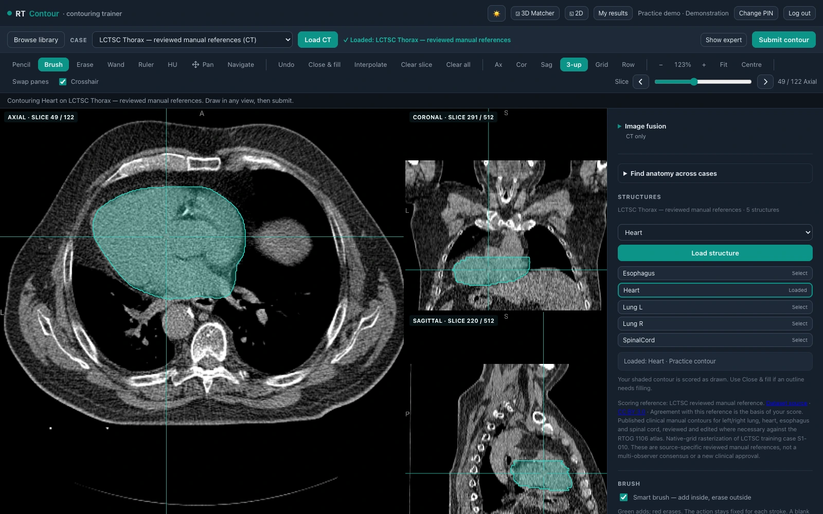
01 / Choose your anatomy
More cases.
More ways to practise.
A growing library brings different anatomies into one workspace. Browse by region, then choose the case and structure for your next attempt.
New cases for your next contouring session.
Explore a lung cancer case with tumour, nodal-target, heart and oesophagus references, two additional prostate planning cases, and two additional pancreatic cancer cases for organs-at-risk practice.
The lung exercise uses one selected 50% breathing phase from the 4D-Lung collection. Pancreatic exercises contain partial small-bowel and stomach/duodenum contours near the treatment region. Review each case description for the available structures and reference scope.
Browse the expanded library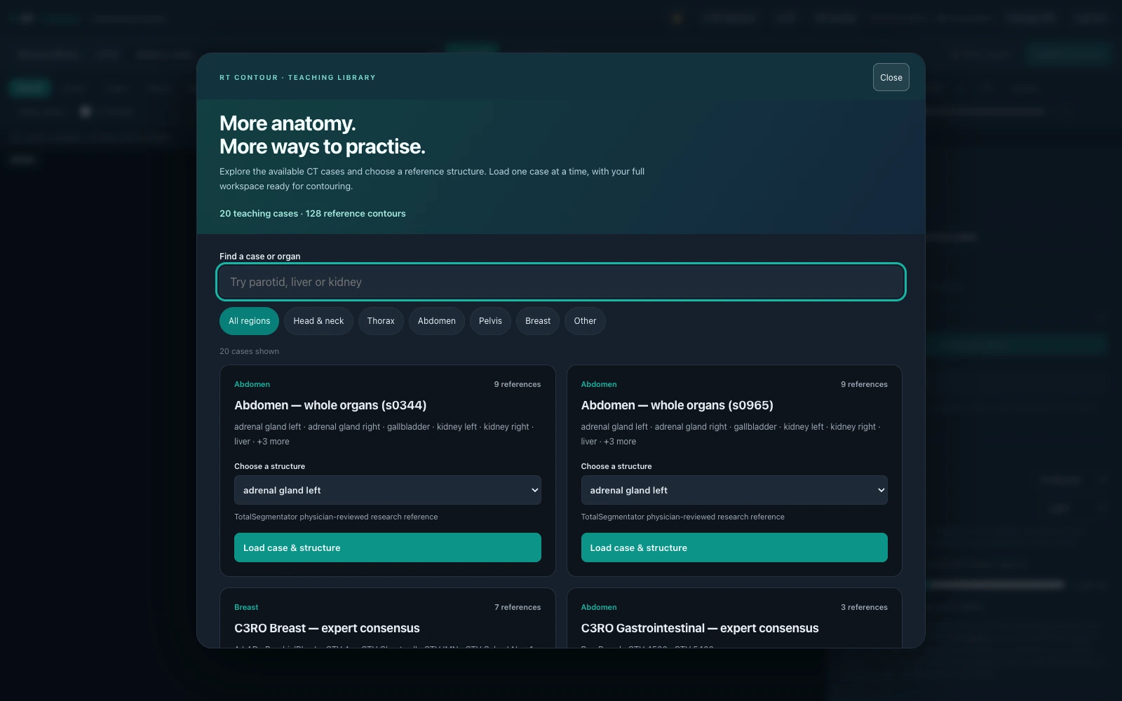
Know what you’re comparing with.
References include published manual contours, research contours, expert-consensus masks and educator-created structures. The app identifies the reference source where available. Some exercises cover part of an organ or a specific subregion; the case notes explain the scope.
About the reference library02 / See the difference
Your contour.
The reference.
The details between.
Submit your attempt to see the regions you share with the reference, the extra areas you included and the areas you missed.
Review the colour overlay and boundary outlines across the scan. Use the recorded metrics to decide what to revisit next.
- Common
- Extra
- Missing
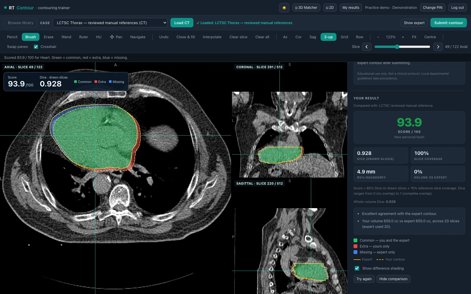
What does the score measure?
The practice score combines 85% Dice overlap on drawn slices and 15% reference slice coverage. The contour you submit is the contour scored; submission does not silently fill gaps.
- Dice overlap
- How much your contour overlaps the reference. Drawn-slice and whole-volume Dice are reported separately.
- Slice coverage
- How much of the reference’s slice extent you attempted.
- Boundary distance
- 95th-percentile boundary distance in millimetres, when available. Large comparisons may defer this metric.
- Volume difference
- The difference between your contoured volume and the reference volume.
These measures support educational comparison. A score or Dice threshold does not establish clinical acceptability.
03 / Revisit your practice
A clearer view
of your own progress.
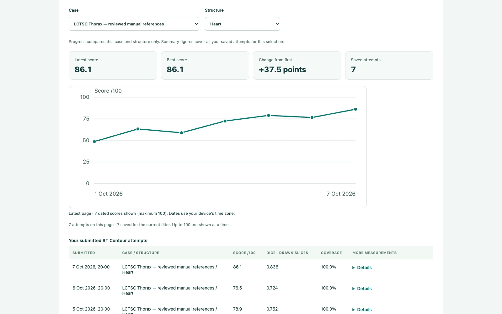
Your saved attempts stay in My results. Return to the same case and structure to review your latest score, your best attempt and how your recorded results have changed.
The tools behind the practice
Work through the anatomy.
Keep control of the contour.
Point or freehand drawing
Place points for a smooth closed outline, or draw freehand. Refine an edge with the brush and eraser.
Assistance you can adjust
Use stroke smoothing, density-aware tools and previewed between-slice interpolation, with Undo for edits.
Linked anatomical views
Inspect axial, coronal and sagittal planes. Pan, zoom, change slices and adjust window and level.
Additional imaging
Where a case includes another scan, blend the images and explore alignment in the same workspace.
Desktop and touch layouts
Open the workspace in your browser. Touch controls keep case selection and tools within reach.
Your own attempt history
Review recorded scores and metrics by case and structure, with a dated graph and detailed table.
Real sources. Clearly described references.
The library includes published teaching and research imaging alongside educator-supplied cases. Manual contours and expert-consensus references are identified separately, with source and licence information available in the app where provided.
RT Contour is an educational practice environment. Case inclusion and technical checks do not establish clinical validation or approval for patient care. Use the case description and your educator’s guidance when interpreting a reference.
Scan screenshots: Yang et al. (2017), Lung CT Segmentation Challenge (LCTSC), v3, The Cancer Imaging Archive, source case S1-010. CC BY 3.0. Teaching adaptation with an illustrative practice contour. TCIA data-use policy: do not identify or contact participants.
Your next attempt starts here
Choose a case.
Make it your practice.
Shared Radiotherapy Education sign-in · Module access required